Musculoskeletal Radiology and Intervention
Musculoskeletal (MSK) Imaging is a subspecialty of radiology that provides diagnostic imaging examinations of the bones, ligaments, muscles and joints. Specifically, musculoskeletal imaging is used to diagnose conditions that include arthropathies, metabolic bone disorders, sports injuries, traumatic injuries, stress related injuries, congenital anomalies, and tumors. Examinations include radiographs, ultrasounds, MRIs, and CTs; procedures include joint aspiration, joint injections, arthrography, and biopsies.
MCR MSK Radiologists are local experts who work closely with orthopedic surgeons, podiatrists, rheumatologists, spine surgeons, as well as general practitioners to provide the best care possible. For example, MCR MSK Radiologists helped establish a monthly Hampton Roads Orthopedics - Sports Medicine - Radiology Interdisciplinary Conference that brings local providers together to discuss best practices in the realm of Sports Medicine and other orthopedic topics. And as the sole Hampton Roads Radiology group teaching Eastern Virginia Medical School (EVMS) Radiology Residents, our subspecialized proficiency is passed on to the next generation of radiologists.
At MCR, we understand that these procedures can be both intimidating and confusing. Our physicians and staff are always willing to answer all questions and make the experience as comfortable as possible.

MRI
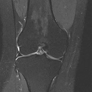
MRI (Magnetic Resonance Imaging) is used in musculoskeletal examinations because of the particular usefulness in evaluating soft tissue structures such as cartilage, muscles, bone marrow, nerves and vessels.
For more general information about the use of MRI in MSK Radiology, please see https://www.radiologyinfo.org/en/info/muscmr structures.
MRI Arthrography
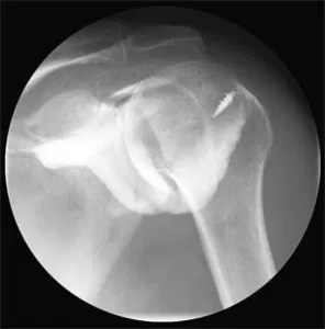
MRI arthrography is a two-step examination, an arthrogram and an MRI exam. It is performed because of its exceptional diagnostic accuracy when there is a need to examine the labrum of the hip and shoulder, as well as certain small ligaments, in particular, of the wrist. MRI Arthrography is also used for complex cases such as postoperative knee and postoperative shoulder evaluations. Prior to the MRI exam, an arthrogram is performed in which a contrast agent is injected into a joint using fluoroscopy for guidance. The contrast allows better visualization of the joint and possible tears of tendons or cartilage on the images produced by the MRI exam. After the contrast has been injected into the joint, the MRI exam is performed. This method of imaging produces highly detailed images.
Computed Tomography
Computed Tomography (CT) examinations are used in musculoskeletal examinations because of their usefulness in evaluating the osseous structures for fractures, healing processes, osseous alignment, infections, and bone tumors. It is also particularly helpful in providing three-dimensional image reconstructions that can be helpful in surgical planning. To help visualize the vascularity of the structures being examined, a contrast agent may be administered.

CT Arthrography
Similar to MRI arthrography, CT arthrography is also a two-step examination, an arthrogram and a CT examination. It is used because of its ability to produce finely detailed images of the osseous structures. Prior to the CT examination, an arthrogram is performed in which a contrast agent (dye) is injected into the joint using fluoroscopy for guidance. After the dye has been injected, the CT exam is performed. The exam generates different views of the osseous structures and together with the contrast, produces highly detailed images.
Joint Aspiration
Joints like the shoulder, hip and knee contain synovial tissue that produces a lubricant like fluid. Joint aspiration is performed when there is a need to remove this fluid to determine the causes of swelling, such as infection or arthritis. First, the skin is numbed adequately. Next, using fluoroscopy or ultrasound for guidance, a needle is placed into the joint and the fluid is aspirated (withdrawn). The fluid is then sent to a laboratory for analysis.
Joint Injection
Joint injection with steroids and analgesics is performed to both confirm the joint as the source of pain and to treat the pain. First, the skin is numbed adequately. Next, using fluoroscopy or ultrasound for guidance, a needle is placed into the joint and the medication is injected.

Sonography
Several of our musculoskeletal radiologists have additional specialty training in musculoskeletal sonography, enabling us to perform diagnostic and interventional ultrasound exams of all joints. Tumors, cysts, and abscesses can be evaluated and aspirated or biopsied if needed.
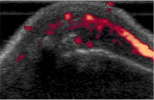
Sonography is an excellent tool for evaluating various conditions such as rheumatoid arthritis, aiding in detection of disease activity and response to therapy. Sonography can also be used to evaluate the anatomy and function of bones and joints using by using direct observation of motion with real-time imaging.


