Body Imaging
High-Quality Diagnostic Body Imaging
Body imaging is a subspecialty of radiology that provides diagnostic imaging examinations of the chest, abdomen, reproductive and gastrointestinal areas. Specifically, body imaging is used to diagnose conditions that include cancer, traumatic injuries, congenital anomalies, infection and inflammation. These examinations include magnetic resonance imaging (MRI), computed tomography (CT), radiographs and ultrasounds of the body, including the abdomen, pelvis and chest. Learn more about Medical Center Radiologists’ comprehensive body imaging services below.
Body Imaging Services In Hampton Roads, VA
MCR’s body imaging subspecialists are fellowship trained to perform a variety of exams. Body imaging can help diagnose and stage cancer as well as a variety of other conditions and diseases. Our body radiologists are experts in body imaging and will work with your physician to determine the preferred imaging method based on the part of the body being examined—if it is a bone, organ or soft tissue—the information needed for a diagnosis and other factors.
Prostate MRI (Screening And Staging Examinations)
Our body radiologists use MRI to take detailed images of the prostate in order to get more details about the cancer, such as where it is, how big it is and if it has spread beyond the prostate. This information helps your physicians make more informed decisions about how to best treat your type of cancer. Learn more about prostate MRI.
MR Enterography (For Inflammatory Bowel Disease)
Using a magnetic field, magnetic resonance (MR) enterography takes highly accurate images of your organs, specifically the small intestine. It can be used to diagnose bleeding or obstruction associated with inflammatory bowel disease with the added benefit of not using radiation. Learn more about magnetic resonance (MR) enterography for inflammatory bowl disease.
Dynamic Pelvic Floor MRI Evaluation
Pelvic floor MRI takes detailed pictures of the pelvic floor, or the muscles that support the pelvic organ. Our body radiologists use these images to assess potential problems with the bladder, vagina, cervix, urethra, uterus or rectum. Learn more about dynamic pelvic floor MRI evaluation.
CT Colonography
A CT colonography is a virtual colonoscopy that uses X-rays to help identify polyps (cancers) in the large intestine. To obtain these pictures, a physician inserts both air and a tube into the rectum to inflate the colon. The goal is to catch polyps early, making CT colonography an important part of early detection of colon cancer. Learn more about CT colonography.
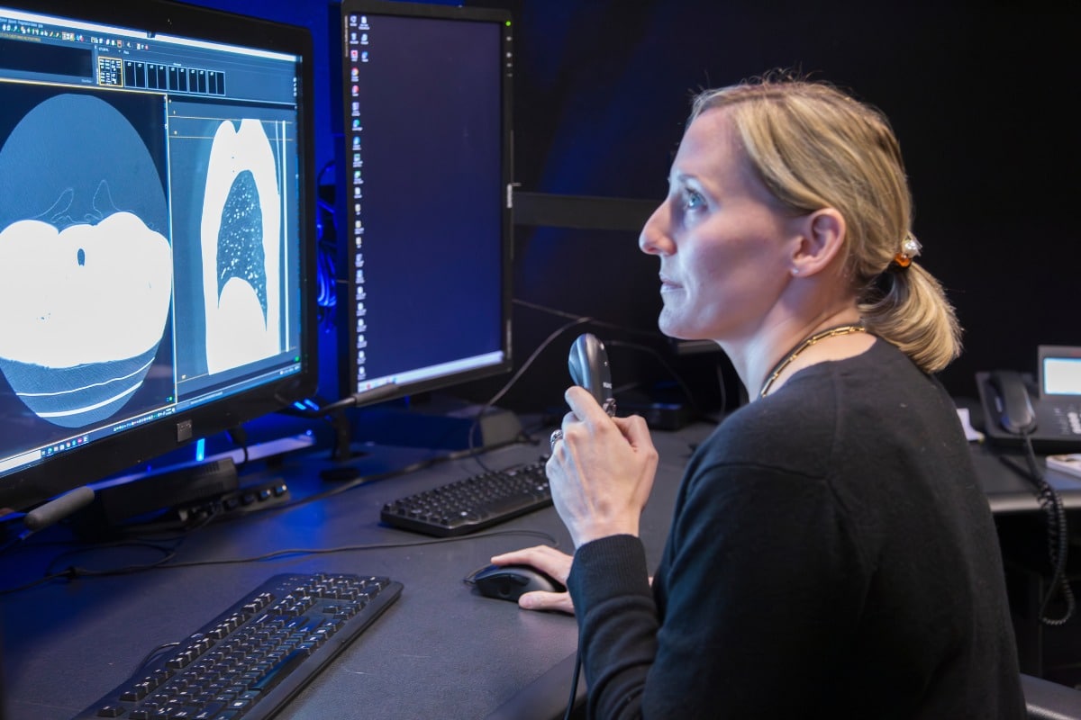
Lung Cancer Screening CT
Lung cancer screening aims to catch lung cancer at its earliest stage in order to give the patient the best possible outcomes. It typically involves blood tests and genetic tests in addition to imaging. However, patients with a high risk of developing lung cancer may opt for a chest scan called low-dose computed tomography (LDCT). LDCT uses cutting-edge technology to take detailed images of the chest. Compared to a traditional chest X-ray, it also uses less radiation. It can help detect cancer in patients who exhibit no other symptoms of lung disease but have a family history or other risk factors. Find more information about lung cancer screening CT.
Additional Body Imaging Services At Medical Center Radiologists
Our body radiologists are uniquely qualified to look for possible abnormalities in the body using non-invasive imaging methods. In addition to the services above, our body radiologists perform the following:
Our Locations