BREAST IMAGING
Mammography, Breast Biopsy And More
At Medical Center Radiologists, we offer breast imaging such as diagnostic and screening mammograms, breast biopsies and more. No matter your breast imaging needs, our experienced team is here to provide you with compassionate, expert care in a comfortable and private setting. Our team offers breast imaging services at the Comprehensive Breast Centers at Sentara Norfolk General Hospital, Sentara Leigh Hospital and Bon Secours Health Center at Harbour View. We offer diagnostic and screening mammograms at Sentara Greenbrier Advanced Imaging Center and screening mammograms at Sentara Independence Advanced Imaging Center.
Available Breast Imaging Services
For a comprehensive list of the breast imaging services we provide, please see the list below, and click on the corresponding link to read more information about each breast imaging service.
Imaging Procedures
Interventional Procedures
Mammograms
To schedule a mammogram at a center staffed by MCR mammographers, choose a convenient location:
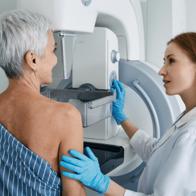
What Is Mammography?
Mammography is a specialized breast imaging technique that uses low-dose X-rays to examine breast tissue. It is widely recognized as the most reliable method for detecting breast cancer at an early stage. In many cases, mammograms can reveal subtle changes in the breast up to two years before they can be physically felt, which greatly improves the chances of successful treatment.
At Medical Center Radiologists, we use digital mammography, also called full-field digital mammography. This advanced technology produces high-resolution images that can be adjusted for brightness, contrast, zoom and orientation, allowing for detailed evaluation on a computer screen.
We also use Computer-Aided Detection (CAD). This system scans mammographic images to identify areas that may need closer inspection—such as unusual densities, masses, or tiny calcium deposits—by highlighting them for the radiologist’s review.
Benefits Of Digital Mammography And CAD
When it comes to breast imaging, there are many benefits to using digital mammography and CAD. Some of these main benefits include:
FAQs About Mammograms
At MCR, we want your breast imaging appointment to be transparent and easy for you to navigate. Below, we have outlined some frequently asked questions about mammography to help you feel prepared for your mammography appointment.
What Is A Breast Biopsy?
This medical procedure takes a small sample of breast tissue to be examined under a microscope. This is typically recommended when breast imaging tests, such as mammograms, ultrasounds, or MRIs, reveal abnormalities, or if there are noticeable changes in the breast or nipple, such as lumps, skin dimpling, scaling or unusual discharge.
Types Of Breast Biopsy Procedures
During your breast biopsy, we will use local anesthesia to numb the area and to make you as comfortable as possible during the procedure. The method we choose depends on the size
Breast Imaging You Can Trust
of the abnormal tissue. Common types of breast biopsy include:
What Happens After A Breast Biopsy?
After the tissue sample is collected, it is sent to a pathology lab, where it is examined for signs of cancer. The pathology report will determine whether the lump is benign, also known as non-cancerous, or malignant, also known as cancerous, and identify the specific type of breast cancer, if present. Based on the findings, your healthcare provider will recommend the most appropriate treatment strategy.
Our Locations